 Dr Todd Born ND reviews current understandings of this increasingly common problem. The definition and classification of chronic kidney disease (CKD) guidelines were introduced by the National Kidney Foundation (NKF) Kidney Disease Outcomes Quality Initiative (KDOQI) in 2002, and were subsequently adopted with minor modifications by the international guideline group Kidney Disease Improving Global Outcomes (KDIGO) in 2004.[1]
These CKD guidelines shifted the concept of kidney disease from that of an uncommon life-threatening condition requiring care by nephrologists to that of a common condition with a range of severity meriting attention by general internists, and demanding strategies for prevention, early detection, and management.[2] The guidelines had a major effect on clinical practice, research, and public health, but also generated substantial controversy.[3]
Chronic kidney disease (CKD) is a worldwide public health problem. In the United States, the prevalence of end-stage renal disease (ESRD) is increasing.[4] The number of patients enrolled in the ESRD Medicare-funded program has increased from approximately 10,000 beneficiaries in 1973, to 86,354 in 1983, and to 615,899 as of December 31, 2011.[5]
Patients with ESRD consume a disproportionate share of healthcare resources. The total cost of the ESRD program in the US was approximately $49.3 billion in 2011. Medicare costs per person per year were more than $75,000 overall, ranging from $32,922 for transplant patients to $87,945 for those receiving hemodialysis therapy.[6]
Data from the UK Renal Registry indicate that there were 41,776 adult patients alive on renal replacement therapy (RRT) in the UK at the end of 2005, a prevalence for adults of 694 per million population (pmp). Information relating to the UK population prevalence of stage 3–5 CKD comes from a large primary care study (practice population 162,113) suggesting an age standardised prevalence of stage 3–5 CKD of 8.5% (10.6% in females and 5.8% in males).[7]
According to a 2009 study in the Journal of Epidemiology and Community Health[8], the overall prevalence of CKD stages 1–5, in England were 14% in males and 13% in females. Only 1.5% of males and 1.3% of females reported being told by a doctor they had CKD. If you are really interested in reading more about CKD in the UK, in 2008 the Royal College of Physicians published “Chronic Kidney Disease, National clinical guideline for early identification and management in adults in primary and secondary care.”
The National Kidney Foundation Kidney Disease Outcomes Quality Initiative (NKF-K/DOQI) workgroup has defined CKD as the following [1], which has been accepted internationally:[9]
- The presence of markers of kidney damage for three or more months, as defined by structural or functional abnormalities of the kidney, with or without decreased glomerular filtration rate (GFR), that can lead to decreased GFR, manifest by either pathological abnormalities or other markers of kidney damage, including abnormalities in the composition of blood or urine, or abnormalities in imaging tests.
OR
- The presence of GFR <60 mL/min per 1.73 m2 for three or more months with or without other signs of kidney damage, as described above.
Based upon representative samples of the United States population, the following studies have estimated the prevalence of CKD in the general population through measurement of markers of kidney damage such as elevated serum creatinine concentration, decreased estimated GFR (eGFR), and presence of albuminuria, including microalbuminuria.
The purpose of CKD staging is to guide management, including stratification of risk for progression and complications of CKD. Risk stratification is used as a guide to inform appropriate treatments and the intensity of monitoring and patient education.[10] In patients who are diagnosed with CKD using the criteria described above, staging of the CKD is done according to:[11]
- Cause of disease
- Six categories of GFR (G stages)
- Three categories of albuminuria (A stages)
Staging patients with CKD according to cause, GFR, and albuminuria enhances risk stratification for the major complications of CKD.
It can be difficult to ascertain the cause of kidney disease. In clinical practice, CKD is most often discovered as decreased eGFR during the evaluation and management of other medical conditions
GFR — The GFR (G-stages) follow the original CKD classification scheme:[12],[13]
- G1 − GFR >90 mL/min per 1.73 m2
- G2 − GFR 60 to 89 mL/min per 1.73 m2
- G3a − GFR 45 to 59 mL/min per 1.73 m2
- G3b − GFR 30 to 44 mL/min per 1.73 m2
- G4 − GFR 15 to 29 mL/min per 1.73 m2
- G5 − GFR <15 mL/min per 1.73 m2 or treatment by dialysis
In general, the presence of the following risk factors for CKD should provoke formal testing for CKD:[14]
- History of diabetes, cardiovascular disease, hypertension, hyperlipidemia, obesity, metabolic syndrome, smoking, human immunodeficiency virus (HIV) or hepatitis C virus infection, and malignancy
- Family history of kidney disease
- Treatment with potentially nephrotoxic drugs
All patients who have risk factors for CKD should be screened with both a urine test for albumin in a first morning or a random “spot” urine sample and a blood test for creatinine to estimate GFR. The preferred screening strategy for albuminuria is measurement of the urine albumin-to-creatinine ratio in an untimed urinary sample.
Depending upon the presence of particular risk factors, additional testing, such as renal ultrasonography, may be required, for example, in patients with a family history of polycystic kidney disease (PKD). The K/DOQI clinical practice guidelines for CKD, as well as other K/DOQI guidelines, can be accessed through the NKF’s web site (www.kidney.org/professionals/kdoqi/guidelines.cfm).
For people with disabilities, and ongoing mental health challenges, as well or independent of chronic kidney disease, the team at advocates for health have compiled this useful resourse. (http://sunrisehouse.com/addiction-demographics/physically-disabled/)
Once the diagnosis is established and the cause and/or potentially reversible factors are identified and treated, CKD should be staged according to the classification proposed by the NKF-K/DOQI Clinical Practice Guidelines for Evaluation, Classification, and Stratification of CKD and subsequently revised.
This classification is based on laboratory evaluation of the severity of kidney disease, association of level of kidney function with complications, and stratification of risks for loss of kidney function and development of cardiovascular disease (CVD). The K/DOQI classification also includes an action plan for each stage of the disease.
The frequency of testing for CKD in high-risk groups has not been rigorously studied, and recommendations are opinion-based. The National Kidney Disease Education Program (NKDEP) of the National Institutes of Health (NIH) recommends yearly testing among diabetic patients.
Hypertensive patients should be tested at diagnosis, upon initiation of therapy, and every three years thereafter. Individuals with a family history of CKD should be tested every three years. Individuals with other risk factors may be tested less frequently.[15]
Although the blood urea nitrogen (BUN) also varies inversely with the GFR, it is generally less useful than the serum creatinine because the BUN can change independently of the GFR.
Because of the problems with changes in creatinine production and secretion, other endogenous compounds have been evaluated in an effort to provide a more accurate estimation of GFR including symmetric dimethylarginine[16] and cystatin C.
The best studied is cystatin C, a low molecular weight protein that is a member of the cystatin superfamily of cysteine protease inhibitors. The serum cystatin C concentration may correlate more closely with the GFR than the serum creatinine concentration.[17] Although cystatin C has been purported to be unaffected by gender, age or muscle mass, higher cystatin C levels have now been associated with male gender, greater height and weight, and higher lean body mass. Cystatin C levels increase sharply with age.[18]
The general management of the patient with CKD involves the following issues:[19]
- Treatment of reversible causes of renal failure
- Preventing or slowing the progression of renal disease
- Treatment of the complications of renal failure
- Adjusting drug dosages when appropriate for the level of eGFR
- Identification and adequate preparation of the patient in whom renal replacement therapy will be required
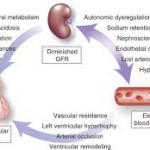
A wide range of disorders may develop as a consequence of the loss of renal function. These include disorders of fluid and electrolyte balance, such as volume overload, hyperkalemia, metabolic acidosis, and hyperphosphatemia, as well as abnormalities related to hormonal or systemic dysfunction, such as anorexia, nausea, vomiting, fatigue, hypertension, anemia, malnutrition, hyperlipidemia, and bone disease. Attention needs to be paid to all of these issues.
Once the patient has reached the stage of near end-stage renal disease (eGFR less than 15 mL/min per 1.73 m2), signs and symptoms related to uremia begin to occur, such as malnutrition, anorexia, nausea, vomiting, fatigue, sexual dysfunction, platelet dysfunction, pericarditis, and neuropathy.
When to send to a specialist? Current guidelines state the following:[20]
- Urine albumin-to-creatinine ratio (ACR) >300 mg/g (34 mg/mmol)
- Haematuria not secondary to urological conditions
- Inability to identify a presumed cause of CKD
- eGFR decline of more than 30% in fewer than four months without an obvious explanation
- Difficult to manage complications such as anemia requiring erythropoietin therapy, and abnormalities of bone and mineral metabolism requiring phosphorus binders or vitamin D preparations
- Serum potassium greater than 5.5 meq/L
- Difficult to manage drug complications
- Patients under the age of 18 years
- Resistant hypertension
There is a nice 2010 article in the Natural Medicine Journal on dietary suggestions in CKD.[21] Regarding fluid intake, “My Doctor Said I Should Drink a Lot! Recommendations for Fluid Intake in Patients with Chronic Kidney Disease” in the Clinical Journal of the American Society of Nephrology is quite helpful.[22]
Protein intake should be adjusted to 0.8 to 1.0 g/kg/day of high biological value protein. Some recommend 0.56 to 0.75 g/kg/day, with 0.35 g/kg/day of high biological value protein. The protein intake is increased by 1.0 g/day of high biological value protein for each gram per day of urinary protein loss. This is performed under close supervision and dietary counseling.[23],[24]
The following nutrients have proven efficacy in the treatment of CKD, ESRD and their respective co-morbidities.
- L-carnitine: 500-2,000 mg daily[25]
- Essential or branched-chain amino acids[26]
- Folic acid: 10 mg daily[27]
- Vitamin B12: 1000 mcg, once a week[28]
- Thiamin: 50-200 mg daily[29]
- Zinc: 50 mg daily[30]
- Balance with copper at this dose: About 3 mg daily
- Vitamin C: 50-100 mg daily[31]
- Periodically measure oxalate levels[32]
- N-Acetylcysteine: 600 mg twice daily[33]
- Omega 3 essential fatty acids: 1.8 g/day of EPA + 1.2 g/day of DHA[34]
- Selenium: 200 mcg three times a week[35]
- Coenzyme Q10: 60 mg three times daily[36]
Botanical medicines:
- Camellia sinensis (decaf green tea)[37]
- Urtica seed[38]
- Rheum palmatum: 1 gram daily[39]
- Lespedeza capilata; Parietaria judaica (1000-1500 mg daily):[40]
- Astragalus:[41] 15 grams daily
- Cordyceps:[42] 1 gram three times daily
- Ganoderma lucidum:[43],[44] 750-1100 mg daily
- Tribulus terrestris:[45]
- Silybum marianum:[46] 140 mg three times daily
- Pre and Probiotics[47]
Exposure to high levels of lead and cadmium have also been shown to have adverse effects on kidney function.[48],[49]
Conclusions
Given the aforementioned information, along with the degree of disease burden in industrialized nations and in conjunction with the ramifications of chronic renal disease, CAM therapies can and may play a crucial role in preventing and mitigating the disease processes.
References
[1] Levey AS, et al. Definition and classification of chronic kidney disease: a position statement from Kidney Disease: Improving Global Outcomes (KDIGO). Kidney Int. 2005;67(6):2089. (View Abstract).
[2] Levey AS, et al. Conceptual model of CKD: applications and implications. Am J Kidney Dis. 2009;53(3 Suppl 3):S4. (View Abstract).
[3] Levey AS. Chronic kidney disease. Lancet. 2012;379(9811):165. (View Abstract).
[4] National Kidney Foundation. K/DOQI clinical practice guidelines for chronic kidney disease: evaluation, classification, and stratification. Am J Kidney Dis. 2002;39(2 Suppl 1):S1. (View Abstract).
[5] United States Renal Data System. USRDS 2013 Annual Data Report: Atlas of Chronic Kidney Disease and End-Stage Renal Disease in the United States. National Institutes of Health. National Institute of Diabetes and Digestive and Kidney Diseases. Bethesda, MD, 2013. (View Abstract).
[6] United States Renal Data System. USRDS 2013 Annual Data Report: Atlas of Chronic Kidney Disease and End-Stage Renal Disease in the United States. National Institutes of Health. National Institute of Diabetes and Digestive and Kidney Diseases. Bethesda, MD, 2013. (View Abstract).
[7] Royal College of Physicians. National clinical guideline for early identification and management in adults in primary and secondary care. CHRONIC KIDNEY DISEASE. 2008. (View Abstract).
[8] Roderick P. Prevalence of chronic kidney disease in England: Findings from the 2009 Health Survey for England. J Epidemiol Community Health 2011;65:A12. (View Abstract).
[9] Levey AS, et al. Definition and classification of chronic kidney disease: a position statement from Kidney Disease: Improving Global Outcomes (KDIGO). Kidney Int. 2005;67(6):2089. (View Abstract).
[10] Levey AS, et al. Conceptual model of CKD: applications and implications. Am J Kidney Dis. 2009;53(3 Suppl 3):S4. (View Abstract).
[11] KDIGO. Chapter 1: Definition and classification of CKD. Kidney Int Suppl. 2013;3:19.
[12] KDIGO. Summary of recommendation statements. Kidney Int 2013; 3 (Suppl):5.
[13] National Kidney Foundation. K/DOQI clinical practice guidelines for chronic kidney disease: evaluation, classification, and stratification. Am J Kidney Dis 2002; 39 (Suppl 1):S1.
[14] Levey AS. Chronic kidney disease as a global public health problem: approaches and initiatives – a position statement from Kidney Disease Improving Global Outcomes. Kidney Int. 2007;72(3):247. (View Abstract).
[15] National Kidney Disease Education Program (NKDEP) (View Abstract)
[16] Kielstein JT, et al. Symmetric dimethylarginine (SDMA) as endogenous marker of renal function–a meta-analysis. Nephrol Dial Transplant. 2006;21(9):2446. (View Abstract).
[17] Stevens LA, et al. Estimating GFR using serum cystatin C alone and in combination with serum creatinine: a pooled analysis of 3,418 individuals with CKD. Am J Kidney Dis. 2008;51(3):395. (View Abstract).
[18] Knight EL, et al. Factors influencing serum cystatin C levels other than renal function and the impact on renal function measurement. Kidney Int. 2004;65(4):1416. (View Abstract).
[19] Schieppati A, et al. Pathophysiology and management of chronic kidney disease. In: Primer on Kidney Diseases, Greenberg, A (Ed), Elsevier Saunders, Philadelphia, 2005, p. 444.
[20] Kidney Disease Outcomes Quality Initiative (K/DOQI). K/DOQI clinical practice guidelines on hypertension and antihypertensive agents in chronic kidney disease. Am J Kidney Dis. 2004;43(5 Suppl 1):S1. (View Abstract).
[21] Henderson J. Dietary Considerations in Chronic Kidney Disease. 2010. (View Abstract).
[22] Wenzel UO, et al. My doctor said I should drink a lot! Recommendations for fluid intake in patients with chronic kidney disease. Clin J Am Soc Nephrol. 2006 Mar;1(2):344-6. (View Abstract)
[23] Ahmed, K. Nutritional management of renal disease. In: Primer on Kidney Diseases, Greenberg, A (Ed). Academic Press, San Diego, CA, 1994, p. 289.
[24] Ikizler, IA. Nutrition and kidney disease. In: Primer on Kidney Diseases, Greenberg, A (Ed). Elsevier, Philadelphia, 2005, p. 496.
[25] Wasserstein AG. L-carnitine supplementation in dialysis: treatment in quest of disease. Semin Dial. 2013 Jan-Feb;26(1):11-5. (View Abstract).
[26] Eustace JA, et al. Randomized double-blind trial of oral essential amino acids for dialysis-associated hypoalbuminemia. Kidney Int. 2000 Jun;57(6):2527-38. (View Abstract).
[27] Pronai W, et al. Folic acid supplementation improves erythropoietin response. Nephron. 1995;71(4):395-400. (View Abstract).
[28] Hoffer LJ, et al. Cobalamin dose regimen for maximum homocysteine reduction in end-stage renal disease. Metabolism. 2005 Jun;54(6):835-40. (View Abstract).
[29] Hung SC, et al. Thiamine deficiency and unexplained encephalopathy in hemodialysis and peritoneal dialysis patients. Am J Kidney Dis. 2001;38(5):941-947. (View Abstract)
[30] Mahajan SK, et al. Zinc deficiency: a reversible complication of uremia. Am J Clin Nutr. 1982 Dec;36(6):1177-83. (View Abstract).
[31] Tomson CR, et al. Correction of subclinical ascorbate deficiency in patients receiving dialysis: effects on plasma oxalate, serum cholesterol, and capillary fragility. Clin Chim Acta. 1989 Apr 14;180(3):255-64. (View Abstract).
[32] Pru C, et al. Vitamin C intoxication and hyperoxalemia in chronic hemodialysis patients. Nephron. 1985;39(2):112-6. (View Abstract).
[33] Tepel M, et al. The antioxidant acetylcysteine reduces cardiovascular events in patients with end-stage renal failure: a randomized, controlled trial. Circulation. 2003 Feb 25;107(7):992-5. (View Abstract).
[34] Donadio JV, Jr. A controlled trial of fish oil in IgA nephropathy. Mayo Nephrology Collaborative Group. N Engl J Med. 1994;331(18):1194-1199. (View Abstract).
[35] Iglesias P, et al. Selenium and kidney disease. J Nephrol. 2013 Mar-Apr;26(2):266-72. (View Abstract).
[36] Singh, et al. Randomized, double-blind placebo-controlled trial of coenzyme Q10 in chronic renal failure: discovery of a new role. J of Nutr Environ Med. 2000 Vol. 10 No. 4 pp. 281-288. (View Abstract).
[37] Ribaldo PD, et al. Green tea (Camellia sinensis) attenuates nephropathy by downregulating Nox4 NADPH oxidase in diabetic spontaneously hypertensive rats. J Nutr. 2009 Jan;139(1):96-100. (View Abstract).
[38] Sayhan MB, et al. Protective effect of Urtica dioica L. on renal ischemia/reperfusion injury in rat. J Mol Histol. 2012 Dec;43(6):691-8. (View Abstract).
[39] Sanada H. [Study on the clinical effect of rhubarb on nitrogen-metabolism abnormality due to chronic renal failure and its mechanism]. Nihon Jinzo Gakkai Shi. 1996 Aug;38(8):379-87. [Article in Japanese]. (View Abstract).
[40] Yarnell EL. Botanical medicines used for kidney disease in the United States. Iran J Kidney Dis. 2012 Nov;6(6):407-18. (View Abstract).
[41] Li M, et al. Meta-analysis of the clinical value of Astragalus membranaceus in diabetic nephropathy. J Ethnopharmacol. 2011 Jan 27;133(2):412-9. (View Abstract).
[42] Li LS, et al. [Experimental study on effect of Cordyceps sinensis in ameliorating aminoglycoside induced nephrotoxicity]. Zhongguo Zhong Xi Yi Jie He Za Zhi. 1996 Dec;16(12):733-7. (View Abstract).
[43] Futrakul N, et al. Treatment with vasodilators and crude extract of Ganoderma lucidum suppresses proteinuria in nephrosis with focal segmental glomerulosclerosis. Nephron. 2002;92(3):719-20. (View Abstract.).
[44] Futrakul N, et. al. Ganoderma lucidum suppresses endothelial cell cytotoxicity and proteinuria in persistent proteinuric focal segmental glomerulosclerosis (FSGS) nephrosis. Clin Hemorheol Microcirc. 2004;31(4):267-72. (View Abstract.).
[45] Kamboj P, et al. Effect of aqueous extract of Tribulus terrestris on oxalate-induced oxidative stress in rats. Indian J Nephrol. 2011 Jul;21(3):154-9. (View Abstract).
[46] Fallahzadeh MK, et al. Effect of addition of silymarin to renin-angiotensin system inhibitors on proteinuria in type 2 diabetic patients with overt nephropathy: a randomized, double-blind, placebo-controlled trial. Am J Kidney Dis. 2012 Dec;60(6):896-903. (View Abstract).
[47] Ramezani A. The Gut Microbiome, Kidney Disease, and Targeted Interventions. J Am Soc Nephrol. 2013 Nov 14. (View Abstract).
[48] Lin JL, et al. Environmental lead exposure and progression of chronic renal diseases in patients without diabetes. N Engl J Med 2003;34:277-86. (View Abstract).
[49] Navas-Acien A, et al. Blood Cadmium and Lead and Chronic Kidney Disease in US Adults: A Joint Analysis. Am J Epidemiol. 2009 Nov 1;170(9):1156-64. (View Abstract).


3 Comments. Leave new
I found this review an interesting read as I have a male client, 35 yrs old, diagnosed with IgAN in 2007. He is a triathlete hoping to undretake an ironman triathlon distance in September.
He eats a healthy diet but he and his doctor have concerns around his protein requirements and its impact on his kidneys and concerns reference his hyperkelimia. He suffers from proteinuria and hematuria too
His blood tests are normal except for his potassium levels which read as follows:
4.8 mmol/L (3.4-5.0) 12/06/2013
5.7 mmol/L 13/01/2014
5.7 mmol/L 21/01/2014
Current medications:
13/01/2014 Doxycycline Hyclate capsules 100mg (To take one capsule a day starting 2 days before travel and continue for 4 weeks after returning from Pakistan).
06/06/2013 Engerix B Prefilled syringe 20 micrograms/1ml (a pre-filled disposable injection – current repeat).
03/09/2013 Rabies Vaccine Powder and solvent for solution for injection 1ml vial (current repeat).
My question is, how can this man continue his training without harming his kidney function and lowering his potassium levels whilst keeping his protein intake low? Indeed, should he continue to train at this level at all?
All dietary and lifestyle suggestions would be gratefully received.
Thank you in advance.
Helen.
Hello, Helen. Thank you for your inquiry about any helpful interventions you may be able to implement regarding IgA nephropathy.
IgA nephropathy is the most common lesion found to cause primary glomerulonephritis throughout most developed countries of the world. Patients may present at any age, but there is a peak incidence in the second and third decades of life. There is approximately a 2:1 male to female predominance in North American and Western European populations, although this difference is not observed among populations in the Pacific Rim.
• D’Amico G. Natural history of idiopathic IgA nephropathy and factors predictive of disease outcome. Semin Nephrol. 2004;24(3):179. (http://tinyurl.com/q38xpjr).
The presence of IgA nephropathy is confirmed only by kidney biopsy. The pathognomonic finding is observed on immunofluorescence microscopy, which demonstrates prominent, globular deposits of IgA (often accompanied by C3 and IgG) in the mesangium and, to a lesser degree, along the glomerular capillary wall.
A consensus on the pathologic classification of IgA nephropathy has been developed by the International IgA nephropathy Network in collaboration with the Renal Pathology Society. In a retrospective analysis, the following variables correlated with renal outcomes:
• Mesangial hypercellularity
• Segmental glomerulosclerosis
• Endocapillary hypercellularity
• Tubular atrophy/interstitial fibrosis
The predictive value of each of these variables appears to be similar in adults and children.
• Working Group of the International IgA Nephropathy Network and the Renal Pathology Society, et al. The Oxford classification of IgA nephropathy: rationale, clinicopathological correlations, and classification. Kidney Int. 2009;76(5):534. (http://tinyurl.com/pqz5zsk).
• Working Group of the International IgA Nephropathy Network and the Renal Pathology Society, et al. The Oxford IgA nephropathy clinicopathological classification is valid for children as well as adults. Kidney Int. 2010;77(10):921. (http://tinyurl.com/nfsq3mn).
There are two major clinical presentations of IgA nephropathy: gross hematuria, often recurrent, following shortly after an upper respiratory infection or athletic exertion; and persistent asymptomatic microscopic hematuria with or without mild to moderate proteinuria.
• Donadio JV. IgA nephropathy. N Engl J Med. 2002;347(10):738. (http://tinyurl.com/omhk3ca).
Patients with IgA nephropathy who have little or no proteinuria (less than 500 to 1000 mg/day) usually have a low risk of progression. However, progressive proteinuria and renal insufficiency develop in a substantial proportion of patients over the long term. Among patients who develop overt proteinuria and/or an elevated serum creatinine concentration, progression to end-stage renal disease is approximately 15 to 25 percent at 10 years and 20 to 30 percent at 20 years.
• Berthoux F, et al. Predicting the risk for dialysis or death in IgA nephropathy. J Am Soc Nephrol. 2011;22(4):752. (http://tinyurl.com/orfs4y9).
Patients with IgA nephropathy who develop progressive disease typically have one or more of the following clinical or laboratory findings at the time of diagnosis, each of which is a marker for more severe disease:
• Elevated serum creatinine concentration
• Hypertension (>140/90 mmHg)
• Persistent (eg, for more than six months) protein excretion above 1000 mg/day
There are two approaches to the therapy of IgA nephropathy:
• General interventions to slow progression that are not specific to IgA nephropathy, including blood pressure control and, in patients with proteinuria, angiotensin-converting enzyme (ACE) inhibitors or angiotensin II receptor blockers (ARBs).
• Therapy with glucocorticoids with or without other immunosuppressive agents to treat the underlying inflammatory disease.
The general interventions are used in all patients at risk for progression (ie, with proteinuria), while immunosuppressive therapy is used in selected patients.
• KDIGO. KDIGO Clinical Practice Guideline for Glomerulonephritis. Kidney Int Suppl. 2012;2(2):209.
Now to directly answer your questions. The intense training of this individual is really predicated on the current condition of his renal function. He should have a thorough evaluation by a nephrologist and have this discussion with him/her regarding those values, as well as the pros and cons of his high intensity exercise regimens.
Nutritional interventions that have good clinical efficacy include low antigen diet, omega-3 fatty acids and the vitamin E family of tocopherols and tocotrienols.
• Ferri C, et al. Low-antigen-content diet in the treatment of patients with IgA nephropathy. Nephrol Dial Transplant. 1993;8(11):1193. (http://tinyurl.com/oqm45gb).
• Sulikowska B, et al. Effect of 12-month therapy with omega-3 polyunsaturated acids on glomerular filtration response to dopamine in IgA nephropathy. Am J Nephrol. 2004 Sep-Oct;24(5):474-82. (http://tinyurl.com/mkko5tk).
• Dillon JJ. Fish oil therapy for IgA nephropathy: efficacy and interstudy variability. J Am Soc Nephrol. 1997 Nov;8(11):1739-44. (http://tinyurl.com/muv64no).
• Kuemmerle NB, et al. Effects of fish oil and alpha-tocopherol in immunoglobulin A nephropathy in the rat. Pediatr Res. 1998 Jun;43(6):791-7. (http://tinyurl.com/kqvuylb).
• Nowak G, et al. γ-Tocotrienol protects against mitochondrial dysfunction and renal cell death. J Pharmacol Exp Ther. 2012 Feb;340(2):330-8. (http://tinyurl.com/mx942hm).
I hope this helps,
Dr. Born
That’s shocking that there were 615,899 people in 2011 that had ESRD and that it is still a big worldwide public health problem. I didn’t even realize that so many people had CKD and even worse ESRD since that is the end-stage and is dangerous. It would be good to see what treatments are available for people with CKD and how they can avoid falling into ESRD so they can be healthier.